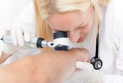
အရေပြားမှန်ဘီလူး ကြည့်ခြင်း
- Examination of the skin by using dermatoscope, which is the high quality magnifying lens with a powerful built-in lighting
- Also called epiluminoscopy or epiluminescent microscopy
- mainly used in pigmented skin lesions by evaluation of colors and microstructures of the epidermis, and papillary dermis
- Accuracy of clinical diagnosis is 60%
Role of dermatoscope
- Easy to diagnose melanoma
- focus on the changes in vascular patterns, vascular structures, colors, follicular abnormalities
- CASH (color, architecture, symmetry, and homogeneity)
Dermoscopy is useful in following case diagnosis:
(1) Melanoma/melanocytic nevi
(2) Basal cell carcinoma
Classical algorithm
- lack of pigment network and the presence of at least one of the following criteria: ulceration, maple-leaf like structure, blue-gray globules, blue-ovoid nests, arborizing vessels and spoke-wheel structures.
Non-classical algorithm
- superficial BCC such as pink-white areas, concentric structures, multiple erosions, multiple in-focus blue-gray dots and fine vessels
(3) Seborrheic keratosis
(4) Squamous cell carcinoma (including Bowen’s disease)
(5) Pityriasis versicolor
Dr. Aye Min Htoo
Dermatologist


Write Reviews
Leave a Comment
No Comments & Reviews